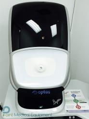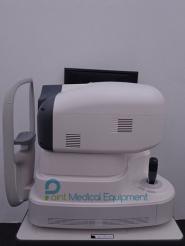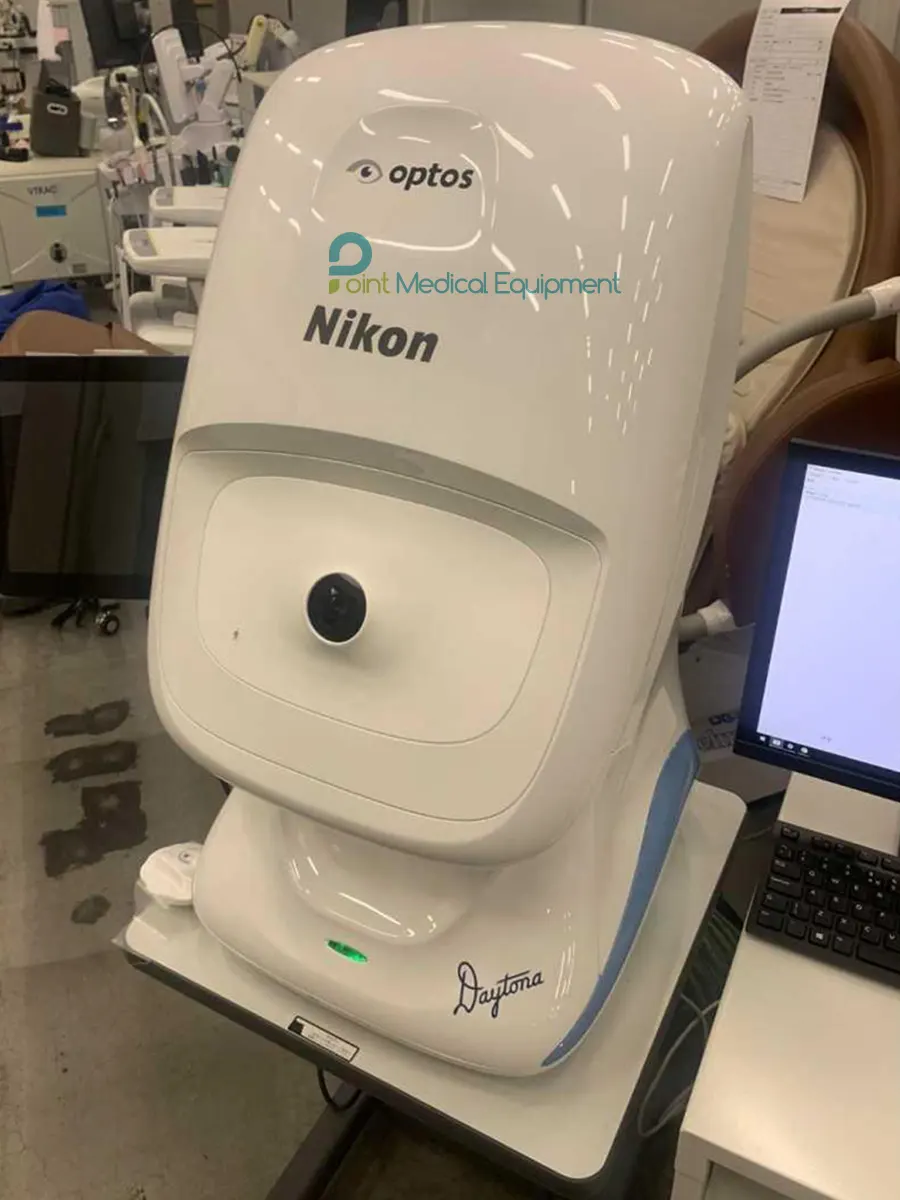
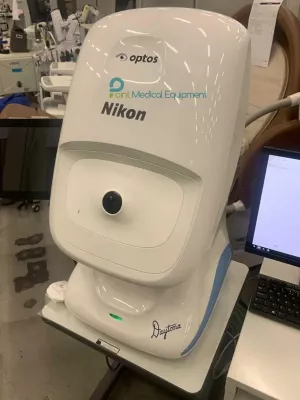
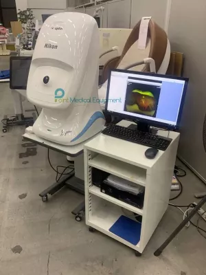
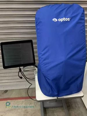
2022 Optos Daytona optomap ultra-widefield retinal imager Package sale includes:
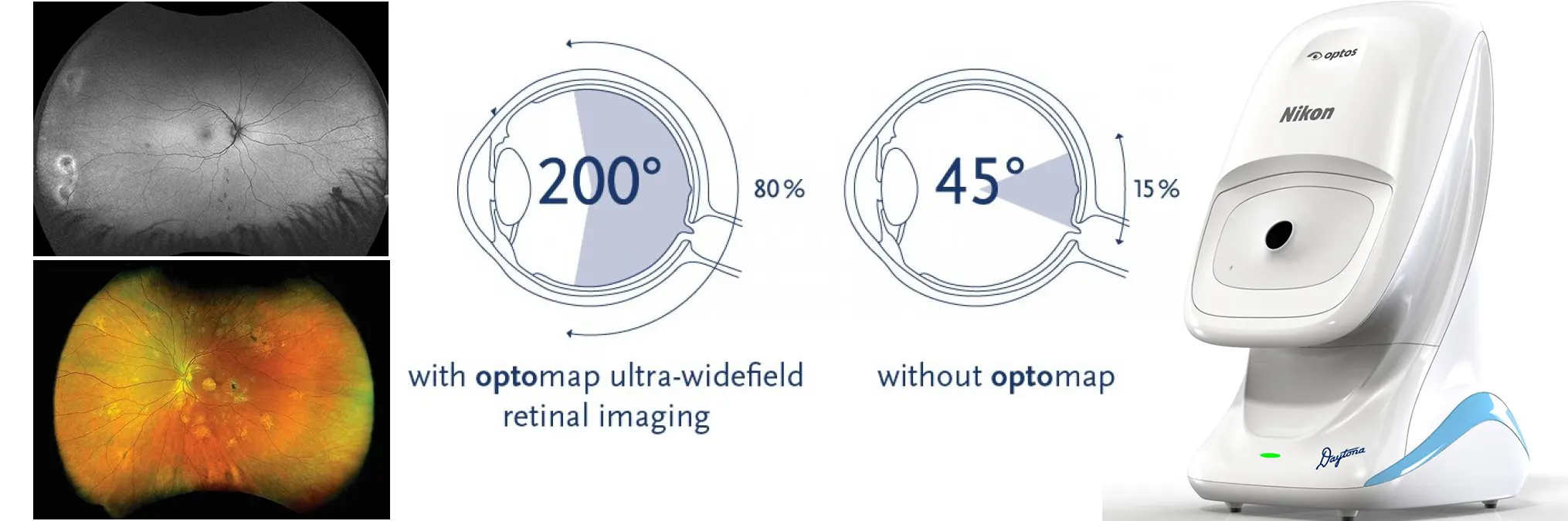
Daytona produces a 200° single-capture optomap® retinal image of unrivaled clarity in less than 1/2 second.
This fast, easy, patient friendly, ultra-widefield (UWFTM) imaging technology was designed for healthy eye screening and has been shown to improve practice flow and patient engagement.
Enhances Clinical Decision-making
Evaluation of the peripheral retina is critical for optimal patient management.
optomap imaging is ideal for peripheral examinations.
Published studies comparing field of view and clinical utility of various widefield imaging systems confirm optomap captures the widest clinically usable field of view and the most retinal pathology.
Improves Practice Efficiency and Economics
Studies show that optomap images are faster to capture and easier to review than traditional patient examination techniques.
A recent study found a 28 minute (33%) reduction in patient visit duration after implementing centralized optomap imaging.
optomap enables practitioners to differentiate their practice and add an additional revenue stream.
OptosAdvance
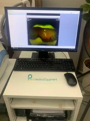
Daytona comes with OptosAdvance an easy to use, browser-based software for documentation, monitoring, and referral processing to facilitate patient management and improve practice flow.
OptosAdvance offers an auto-montage tool to quickly capture and merge a series of images into a single 220° montage showing 97% of the retina.
The software also includes tools for accurate distance and area measurements even in the far periphery.
Features
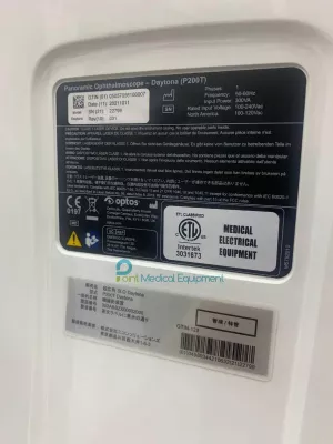
| Daytona P200T optomap UWF Imaging | RESOLUTION optomap: 20 μm, optomap plus : 14 μm |
| LASER WAVELENGTHS Red laser: 635 nm Green laser: 532 nm (for AF) |
|
| EXPOSURE TIME Less than 0.4 seconds |
|
| Daytona P200T System | FOOTPRINT Width: 550 mm / 22 in Depth: 550 mm / 22 in including chinrest Height: 608-632 mm / 24-25 in |
| WEIGHT 34 kg / 75 lbs |
|
| TABLE SPACE REQUIREMENTS (not including wheel position) Width: 887 mm / 35 in Depth: 623 mm / 24 in |
|
| LASER CLASS Laser safety class-1 following EN60825-1: 2007 and 21 CFR1040.10 and 1040.11 |
|
| SYSTEM VOLTAGE US: 100-120V at 50/60Hz, 3A, EU/AU: 200-240V at 50/60Hz, 1.5A |
|
| POWER CONSUMPTION 300VA |
|
| COMMUNICATION PROTOCOL DICOM Compatible |