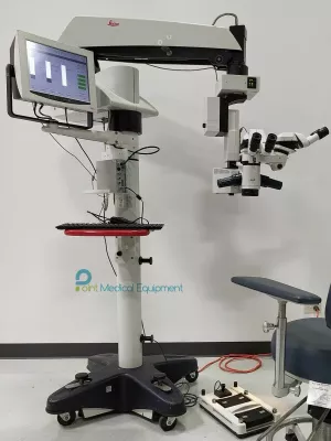

* This system is in very good condition, 6 month warranty.
* 100% functional, the genuine plastic cover still in the stand body.
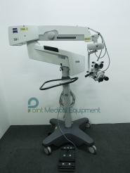
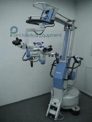
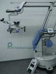
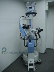
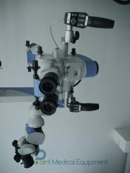
Moller-Wedel Hi-R 1000 Surgical Microscope FS4-20 stand includes:
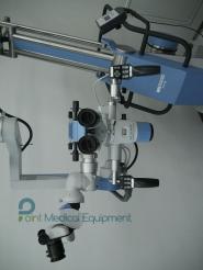
- Hi-R1000 surgical microscope
- Floor stand FS4-20 (built-in xenon light source 300W)
Features:
-Unique drive system SensoServo, which retains the microscope in a balanced position in all the configuration and optical parts.
-Power zoom 1:6
-Diaphragm for protection from blinding light
-Control depth
-RS232 interface for interacting with your navigation device
-Computer control all motorized functions
-Optional: ICG fluorescence and ALA
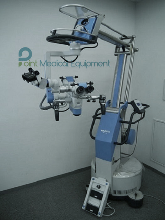
The perfect microscope for neurosurgery Moller Hi R-1000, which is constantly in a balanced State. Floor standing tripod FS 4-20 Moller microscope and Hi-R 1000 SensoServo technology ensures a stable position system even when changing accessories. This high end microscope is useful for micro vascular hand and finger transplants, plastic and reconstructive surgeries.
Features:
PermaBalance | Fly-by-wire technology
The microscope should support the user during his work and not put him under stress. The system MÖLLER 20-1000 follows the ‘fl y-by-wire’ technology, which enables the user to control the complex system with only minimal force. With the support of the programmable SensoGrip, the microscope can easily be positioned, even when draped. Möller’s new SensoServo drive provides precise and quick reaction.
During operation, the assistant may reposition the observer tube without affecting the balance. The brakes and the motor-driven controls are activated via either SensoGrip. While the characteristics of motion are individually set on the control panel, mechanical positioning is performed on the SensoGrip.
The optical properties comply with the highest standards. The apochromatic optics with the correction of residual aberration provides perfect color fi delity, strong contrast, and high resolution. An exceptional depth perception is achieved due to the unique stereo base of 25 mm, enhanced by an iris diaphragm for increased depth of fi eld. The variable objective lens permits working distances from 224 to 510 mm (nominal) without lens exchange. Other distance ranges are available.
Light and Imaging
The base of the fl oor stand houses a powerful 300 W xenon light source. According to your request, the base also contains a back-up lamp module for quick exchange or a second independent 300 W xenon light source.The light is fi ltered against ultraviolet and infrared radiation to protect the surgeon’s eyes and the patient’s tissue. The light intensity can be controlled via hand grip or foot switch. Diaphragms in the microscope head may be selected during the operation to avoid glare
For ALA and/or ICG fl uorescence special light sources are used. They contain fi lters which automatically snap in, when the fl uorescence mode is selected on the hand grip, as do the observation fi lters in the microscope head. A special light guide retains all important light constituencies from the light source to the microscope head.
The arrangement of the heavy light sources in the fl oor stand base provides a low center of gravity thus allowing the system to be slim and unobtrusive in the operating room. The wire connections are also close to the functional parts in the base, easily accessible but well protected behind a cover
MÖLLER MIOS
MIOS stands for Microscope Imaging and Operation System. Its prime functions are recording of operation scenes, capturing and recording of snapshots together with proper identifi cation of patients and hospital data. Images and/or streams can be stored on DVDR/-RW, HDD, USB sticks, external USB hard disks, or transmitted to the hospital PACS system via DICOM
MÖLLER MEDIS
MÖLLER MEDIS (Microscope External Display) turns the operating microscope into a microsurgical image control center. The high-resolution screen, mounted above the oculars, provides images or data for the surgeon which he may see by momentarily looking up from the oculars. The touch screen allows to control numerous functions sterilely through the drape. Three versions are available:
MÖLLER MEDIS M
Control of video recording, snapshots, ICG angiography observation
MÖLLER MEDIS E
As MÖLLER MEDIS M plus connection of endoscopic cameras via video converter
MÖLLER MEDIS I
As MÖLLER MEDIS E plus access to various data
sources via IP addresses