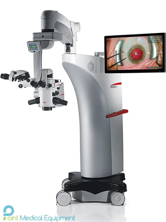
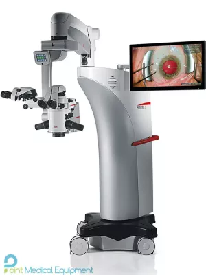
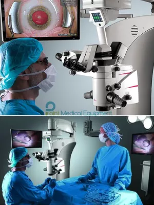
The Proveo 8 ophthalmic microscope provides the exact image you need at each moment of your procedure. Like a precision timepiece every element of the Proveo 8 microscope interconnects and works in perfect synchrony to optimize your view.
Simple to start, fast to finish
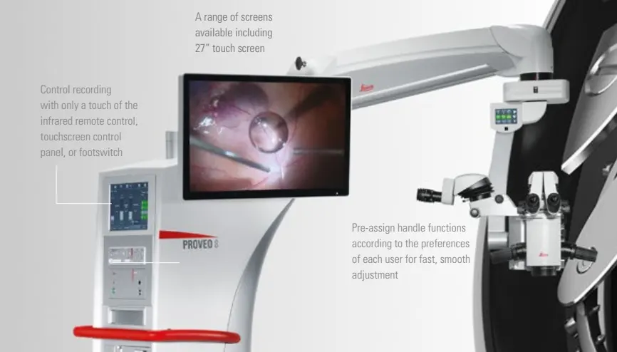
Save precious time between surgeries for yourself and your OR team, with easy preparation and fast transition. The intuitive touch-screen control unit makes set-up easy.
At the end of the surgery simply move the swing arm up and all microscope functions automatically reset and the recorder stops. The microscope is immediately ready for the next case
Ergonomic means efficient
During surgery, your physical well-being can influence your concentration and efficiency.
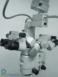
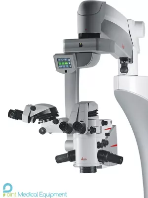
Choose from a large selection of binoculars and three different objective lens types to meet your individual physical requirements and those of your assistant
Smooth, comfortable working
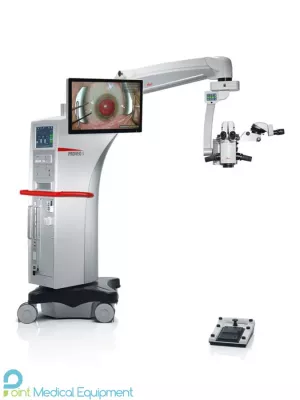
Pre-program the wireless footswitch with key functions and maintain your surgical workflow in a comfortable posture.
Switch functions with just a tap of the foot. Functions available include vitreoretinal (VR) mode, tilting position, quick focus, and diameter of red reflex illumination. Position the footswitch exactly where you need it thanks to the lightweight, cable-free design.
Don’t waste time re-adjusting your microscope during anterior or posterior surgery – work interruption-free with the Proveo 8.
Features:
Leica Microsystems introduces the latest Leica Proveo 8 ophthalmic microscope, which provides exceptional functionality for the Hi-End class of microscopes. Well proven in the M844, the unique 4-channel Leica QuadZoom ™ system is now enhanced with FusionOptics technology to provide the surgeon with an unrivaled depth of field without sacrificing resolution.
The surgeon's completely independent optical channels in combination with excellent optics allow for excellent image quality in any operation and at a sufficiently low light level. The combination of the path of the optical systems of the surgeon and the assistant in one scheme allows to achieve ideal synchronization of the magnification and diameter of the field of view for the surgeon and the assistant. The assistant can individually fine-tune the focus separately from the primary surgeon.
In addition, the optical unit of the microscope is equipped with fast focus and fast tilt functions, which significantly optimize the workflow, allowing you to reduce the time spent on each operation, providing additional comfort for the surgeon.
The microscope is supplied with an EnFocus objective designed for intraoperative OCT, and the Proveo 8 design allows for quick retrofitting of intraoperative OCT in 1 day.
Leica Proveo 8 Ophthalmic Microscope Technical Specifications
TECHNICAL SPECIFICATIONS
| Optics and Illumination | |
| FusionOptics | For increased depth of field and high resolution for main surgeon and assistant |
| OptiChrome optics | For high contrast, high resolution, natural colors without chromatic aberrations |
| Magnification | 6:1 zoom, motorized |
| Total magnification | 4.1× to 24.5× with 10× eyepiece 5.1× to 30.7× with 12.5× eyepiece |
| Focus range | 75 mm |
| Objective / working distance |
WD 175 mm/f = 200 mm , WD 200 mm/f = 225 mm , WD 225 mm/f = 250 mm |
| Field of view | 51.4–8.6 mm Ø with 10× eyepiece |
| Eyepieces | Wide-field eyepieces for persons wearing glasses 8.3×, 10× and 12.5× dioptric adjustment, ± 5 diopter settings, adjustable eyecup |
| Direct illumination with 2 LED lamps |
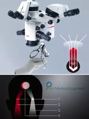
Main light CoAx 4 coaxial illumination > Illumination unit for generating a clear and stable Red Reflex, decreasing stray light through the sclera and increasing the image contrast > Integrated keratascope and slit illumination Conversion filters allow surgeon to select preferred color temperature of main illumination |
| Adjustable CoAx 4 | Diameter of coaxial illumination is adjustable between 4 and 23 mm via footswitch |
| Fine focus | Available for assistant and integrated camera or external 1/3 camera with C-mount interface |
| Upgradeability | |
| OpenArchitecture | Prepared for integration of video camera systems, digital recording and imaging systems such as IOLcompass, EnFocus OCT, and monitors |
| Connectors | > Numerous built-in connectors for video and control data transfer > Internal power supply 12 VDC, 19 VDC, 24 VDC and AC terminals |
| 2D/3D HD Video | Optional fully integrated 2D HD and/or 3D HD video and recording |
| Maneuverability | |
| Optics | > 360° rotation > 15° /+ 105° motorized inclination tilt |
| XY speed | Zoom linked XY speed |
| XY range | 62 × 62 mm |
| Balancing | Adjustable gas spring via balancing knob |
| Brakes | Floor stand with 4 electromagnetic brakes |
| Monitor arm | 860 mm flexible arm with 4 axis for rotation and inclination, max. weight 15 kg and up to 32” |
| Control | |
| Control unit | > User-friendly, individually programmable touch-screen (up to 30 surgeons) for control of motor functions and light intensity > Menu selection based on unique software for user-specific configuration > Built-in electronic auto-diagnosis and user support > Software independent hard keys and indicator for illumination > Data shown by means of LCD |
| Control elements | > Rotary handles > 14-function and 12-function wireless footswitch with optional back-up cable |
| IR sensor | Remote control of the HDR recorder |
| Indicators | > LED for video record status > Surgeon information panel for setting status |
| Construction | |
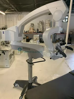
| Floor stand | Four 360° rotating castors (Ø150 mm), parking brake |
| Materials |
> Coated with antimicrobial paint > Conforming with RoHS |
| Load | > Floor stand max. 8.0 kg from microscope dovetail ring interface > C42/CT42 max. 8.0 kg from dovetail ring interface |
| Weight |
> Floor stand approx. 350 kg without load > C42 ceiling mount total approx. 260 kg > CT42 telescope mount total approx. 200 kg |
| Technical data | |
| Power connection | > 1100 VA 50/60 Hz > 100–240 V~ 50/60 Hz > 2 × T10 AH 250 V |
| Protection class | Class 1 |