Leica M530 OHX features include:
Additional Accessories:
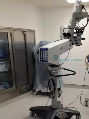
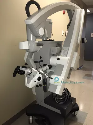
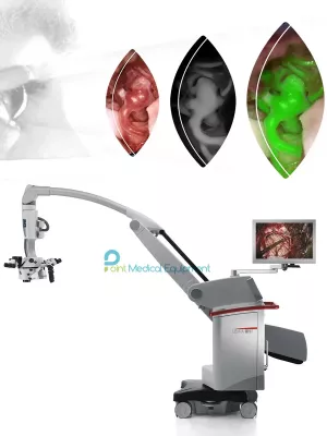
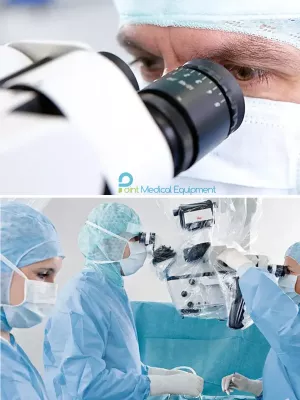
The M530 OHX Neurosurgery Microscope - stay focused through every step of every surgery. Exclusive FusionOptics technology combined with advanced optics and TriFluoro – three integrated fluorescence modes – enable you to focus on every critical detail. The fully ergonomic design helps you and your team to achieve comfortable working positions and avoid physical discomfort. Whether performing delicate neurosurgery, or complex reconstructive microsurgery, you remain focused on delivering optimal results for your patient.
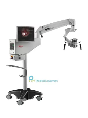
When you require additional visual information, for example from endoscope or IGS system, opt for the CaptiView image injection module.
See more with FusionOptics
FusionOptics, the groundbreaking technology from Leica Microsystems, unites an enhanced depth of field with high resolution to create an optimal view of the surgical field.
A larger area in full focus also means that there is less need to refocus the microscope which helps to streamline workflow:
The Technology of FusionOptics
Images you can rely on
For brilliant images and increased depth perception in deep cavities, the Leica M530 OHX combines FusionOptics technology with world-renowned optical quality and state-of-the-art illumination.
TECHNICAL SPECIFICATIONS
| OPTICS AND ILLUMINATION | |
| FusionOptics | For increased depth of field and high resolution for main surgeon and opposite assistant |
| Fully apochromatic optics | For high contrast, natural colors without chromatic aberrations |
| Magnification | 6:1 zoom, motorized |
| Total magnification | 1.0× to 12.1× with 10× eyepiece |
| Magnification multiplier | 1.4× (optional) |
| Focus | Motorized via multifocal lens, with manual adjustment |
| Fine focus | ±5 diopter available for opposite assistant (ULT) |
| Objective / working distance |
225–600 mm, motorized multifocal lens, continuously adjustable and manual adjustment option |
| Field of view | 17.4 to 210 mm ø with 10× eyepiece |
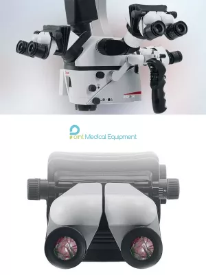
| Eyepieces | Wide-field eyepieces for persons wearing glasses 8.3×, 10× and 12.5× dioptric adjustment, ±5 diopter settings and adjustable eyecup |
| Integrated 360° rotatable adapter |
For main surgeon binocular (IVA, ULT) and opposite assistant (ULT) |
| Illumination | - High-output 2x 400-W redundant xenon arc-lamp systems via fiber optics cable - Continuously variable illumination field diameter with Gaussian distribution - Continuously adjustable brightness at constant color temperature |
| SpeedSpot | Laser focusing aid for fast and exact positioning of the microscope |
| Carrier for monitor | 700 mm flexible arm with 4 axis for rotation and inclination |
| MODULARITY | |
| Basic: IVA530 | Full stereo view for main surgeon, semi stereo view for 2 side assistants and C-mount interface for camera (HD or SD) - Light distribution: 67 % for surgeon, 23 % for side assistant, 20 % for C-mount port |
| Standard: ULT530 | Full stereo view for main surgeon and opposite assistant, semi stereo view for up to 2 side assistants - High sensitivity, built-in IR video camera with 1/2“ CCD - Optional integrated HD Camera (HD C100), FL400, and FL560 - Light distribution: 50 % for main surgeon, either 20 % for each side assistant or 40 % for opposite assistant |
| FL400 | FL400 oncological fluorescence observation filter module |
| FL560 | FL560 investigational fluorescence observation filter module |
| Advanced: FL800 ULT &/or CaptiView image injection |
Full stereo view for main surgeon and opposite assistant, semi stereo view for up to 2 side assistants - CaptiView HD image injection (optional) - FL800 vascular fluorescence with builtin NIR camera (optional) - Optional: C-mount interface for camera (HD or SD) |
| OpenArchitecture | - Easy integration of neuronavigation systems and laser systems - Prepared for integration of video camera system and digital recording system |
| Connectors | - Numerous built-in connectors for video, IGS and control data transfer - Internal power supply 12 VDC, 19 VDC and AC terminals |
| 2D/3D HD Video | Fully integrated 2D HD and/or 3D HD video and recording |
| CONTROL | |
| Control unit | - Programmable touch-screen with userfriendly Graphical User Interface for control of microscope and stand - Built-in electronic auto-diagnosis and user support - Software independent hard keys for illumination and auto-balancing - Indicator for main/backup illumination and fluorescence modes |
| Control elements | - Pistol handle with 10 programmable functions - Optional mouthswitch - Optional 12-function wireless footswitch |
| IR sensor | For remote control of the external HD C100 camera |
| SAFETY | |
| AutoIris | Built-in automatic zoom-synchronized illumination field diameter, with manual override and reset feature |
| BrightCare Plus | Safety function through working distancedependent limitation of the brightness, controlled by a built-in luxmeter |
| CONSTRUCTION | |
| Base | 690 × 690 mm with four 360° rotating casters with a diameter of 150 mm each, one parking brake |
| Materials | All solid metal construction coated with antimicrobial paint |
| Load | Min. 6.7 kg, max. 12.2 kg from microscope dovetail ring interface |
| Weight | Approx. 320 kg without load |
| Indicator | LEDs for fluorescence mode status and video record status |
| TECHNICAL DATA | |
| Ambient conditions in use |
- +10 °C to +40 °C - +50 °F to +104 °F - 30 % to 95 % rel. humidity - 800 mbar to 1060 mbar atmospheric pressure |
| Power connection | - 1600 VA 50/60 Hz - 100 V, 120 V, 220 V, 240 V (+10 %/–15 %) - 2 × T10 AL 100/120 V - 2 × T8 AL 220/240 V |
| Protection class | Class 1 |