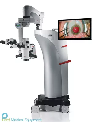
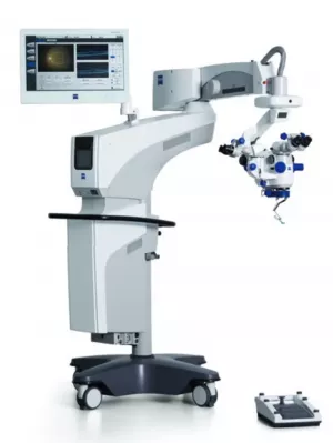
ZEISS OPMI LUMERA 700 Seeing to succeed with the microscope for every ophthalmic specialty, whether preserving or restoring a patient’s sight, the OPMI LUMERA 700 from ZEISS is the surgical microscope for every ophthalmic specialty. Experience markerless IOL alignment and integrated intraoperative OCT3 imaging – all in one device – from the ophthalmic microscope market leader.
OPMI LUMERA 700 offers a true revolution in lighting quality with an intuitive and versatile instrument and It has a built-in optical coherence tomography (OCT) that allows non-invasive insight into the deep layers and internal structures of the eye.
ZEISS OPMI LUMERA 700 is part of our commitment to helping you succeed in your OR. It’s also part of the ZEISS Cataract Suite, which includes leading products designed to work together for markerless toric IOL alignment.
Seeing to succeed in cataract surgery Precise* and efficient** markerless toric IOL alignment. With ZEISS CALLISTO eye markerless alignment, manual marking steps can be skipped altogether for an efficient and precise* toric IOL alignment to reduce residual astigmatism. For cataract surgeries, ZEISS OPMI LUMERA 700, with its well-known patented SCI illumination, ZEISS optics and CALLISTO eye ® from ZEISS provides the best anterior views and precise* assistance functions.
”I save 6 minutes per patient and improve alignment precision by 40% compared to manual marking. Wolfgang Mayer, MD, Augenklinik der Universität München, Germany”
Cataract assistance functions for every step of the surgery The assistance functions of ZEISS CALLISTO eye are completely surgeon-controlled – with either the foot control panel or handgrips.
 |
 |
 |
 |
 |
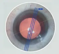
| Z ALIGN Perform toric IOL centration on the visual axis provided by the IOLMaster and perform rotational alignmen |
Incision Position incisions, optionally on the steep axis; add opposite clear cornea incision and paracenteses |
Rhexis Precisely* size and shape capsulorhexis and align the IOL on the visual axis provided by the IOLMaster |
LRI Perform relaxing incisions |
K TRACK Estimate the local corneal curvature in combination with a keratoscope |
 |
||||
Efficient markerless IOL alignment
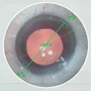
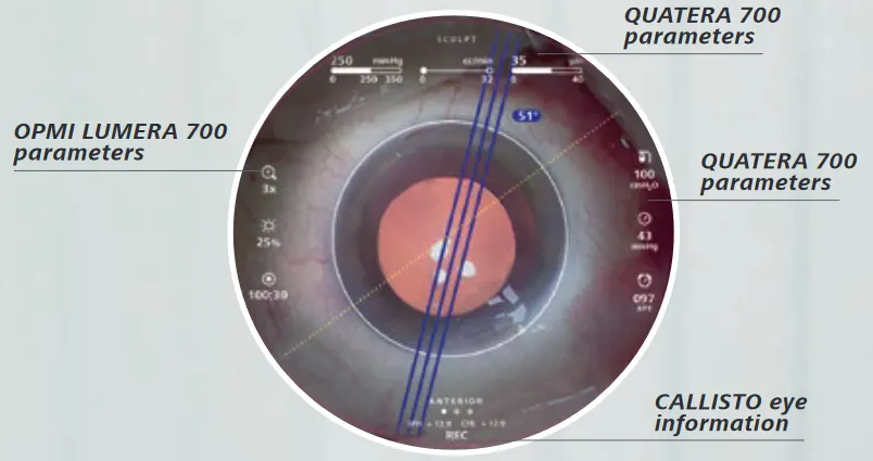
Starting with a biometry reference image from the IOLMaster from ZEISS, data is transferred smoothly to CALLISTO eye. This data is used to create overlays in the eyepiece. Save time, increase efficiency and reduce residual astigmatism when you:
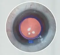
Data from QUATERA 700 from ZEISS is integrated in the visual field of the ocular and screen for a better overview
Efficient surgery setup
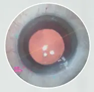
The image quality check supports you to optimize light intensity, magnification and centration of the microscope to efficiently set up the reference axis. The well-proven* eye tracking automatically compensates for eye movements and supports the use of the assistance functions
”CALLISTO eye enabled easy and exact toric IOL alignment in all cases.” Prof. Findl, VIROS, Hanusch Hospital, Vienna, Austria
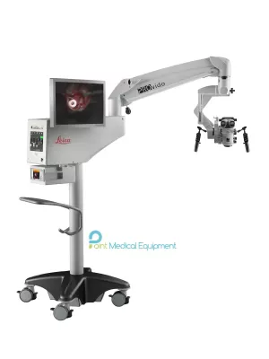
OPMI LUMERA 700 operating microscope from ZEISS is an intuitive and versatile instrument and its technical equipment is among the absolute top. It has a built-in optical coherence tomography (OCT) that allows non-invasive insight into the deep layers and internal structures of the eye. This information is transmitted using a color scale to a three-dimensional 3D image.
With the features integrated into the OPMI Lumera 700, you are optimally prepared for anything: not only for cataract and retinal surgery the next day but also for the future. The system is extremely versatile and, with the appropriate accessories, gives you exactly the tool you need for both anterior and posterior segments.
The OPMI Lumera® 700 offers a true revolution in lighting quality. With Stereo Coaxial Illumination (SCI™). You take advantage of unprecedented recognition of details. The kit includes 0° co-observation with independent magnification and focus control and an image inverter built into the binoculars.
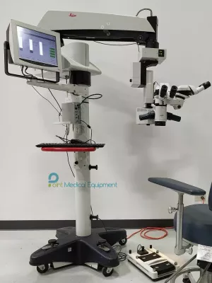
OPMI Lumera 700 is an excellent specialized surgical microscope for all areas of ophthalmic surgery. This provides:
Carl Zeiss Opmi Lumera 700 is an ophthalmic operating microscope for retinal and glaucoma surgery, cataract and corneal pathologies. Equipped with the integrated Carl Zeiss Rescan 700 OCT imaging system, which allows you to visualize the structures of the eye to the smallest detail and in the correct physiological form. OPMI LUMERA 700 is used for several types of operations – gray and green cataracts, and corneal and retinal operations.
WHAT MAKES THE LUMERA OPMI 700 UNIQUE
Areas of use
Cataract treatment
The Carl Zeiss Opmi Lumera 700 provides an unrivaled view of the anterior segment of the eye with patented SCI illumination, Carl Zeiss optics, and the Carl Zeiss Callisto Eye system. The device is equipped with an IOL alignment function for efficient and more accurate alignment of a toric IOL with lower residual astigmatism.
Glaucoma surgery
The Carl Zeiss Opmi Lumera 700 reduces cell loss, reduces handling time, and helps the surgeon make quick decisions during surgery. Carl Zeiss Rescan 700 in glaucoma surgery and canaloplasty procedures helps in monitoring stent implants in hard-to-see places.
Treatment of pathologies of the cornea
The built-in intraoperative OCT allows visualization of the physiological shape of the cornea in two different scanning modes.
Retinal surgery
When performing retinal surgery, the Carl Zeiss Opmi Lumera 700 microscope provides excellent clarity and a good field of view, as well as an unrivaled view of the anterior segment of the eye, thanks to the patented SCI illumination, Carl Zeiss optics, and the Carl Zeiss Callisto Eye system.
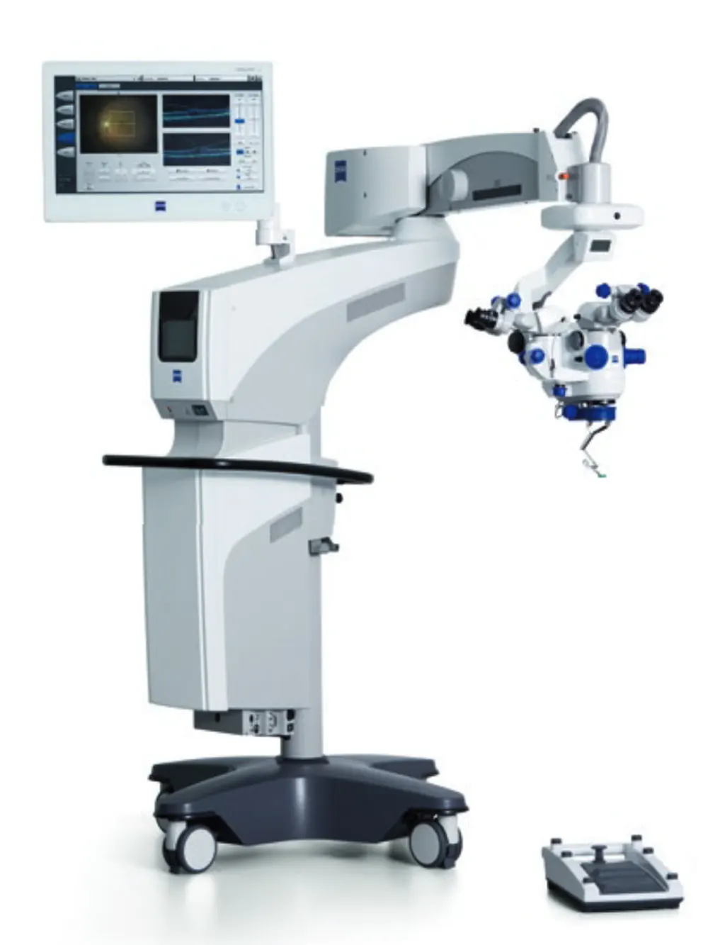
OPMI LUMERA 700 from ZEISS TECHNICAL DATA
| ZEISS OPMI LUMERA 700 | |
| Surgical microscope | Motorized zoom system with apochromatic lens, zoom ratio 1:6 |
| Magnification factor = 0.4 x – 2.4 x | |
| Focusing: electric / motorized, focus range: 70 mm | |
| Objective lens: f = 200 mm (optionally also f = 175 mm or f = 225 mm with support ring | |
| Binocular tube: Invertertube E (optionally also Invertertube, 180° swivel tube, f = 170 mm, inclined tube, f = 170 mm | |
| Wide-angle eyepiece 10 x (optionally also 12.5 x) | |
| Light source | SCI: Coaxial and full-field illumination |
| Fiber-optic illumination Superlux ® Eye: - Xenon short arc reflector lamp with HaMode filter - Backup lamp in lamp housing, can be slid into position manually |
|
| LED fiber-optic illumination: - Near-daylight color temperature - 50,000 hour lifetime at 50% light intensity - HaMode filter - 25% gray filter |
|
| For all light sources: - Blue blocking filter - Optional: Fluorescence filter |
|
| Integrated slit illuminator | Slit widths: 0.2 mm, 2 mm, 3 mm, 4 mm Slit height: 12 mm |
| XY coupling | Travel range: max. 61 mm x 61 mm Automatic centering at the touch of a button |
| Video monitor | 23.6” LCD display Resolution: 1,928 x 1,080 |
| Stand | Maximum permissible weight load of the spring arm: When the surgical microscope is attached to the arm (without tube, eyepiece or objective lens) and the XY coupling is also attached, a maximum of 9 kg of additional accessories can be attached to the spring arm |
| ZEISS intraoperative OCT | |
| OCT engine | SD (spectral domain) OCT Wavelength 840 nm Scanning speed 27,000 A-scans per second |
| Scan parameters | A-scan depth: 2.9 and 5.8 mm in tissue Axial resolution: 5.5 μm in tissue Scan length adjustable 3–16 mm Scan rotation adjustable 360° Scan modes for live and capture acquisition Live: 1-line, 5-lines, cross hair Capture: 1-line, 5-lines, cube |
| ZEISS RESIGHT family | |
| Mechanical data | Focus range with LH175 lens holder: 31 mm (position of intermediate image) |
| Focus range with LH200 lens holder: 38 mm (position of intermediate image) | |
| Rotation angle of lens revolver and holder: 0°–360° | |
| Lenses included | 60D, 128D |
| Weight | ZEISS RESIGHT 500 (manual): 0.45 kg ZEISS RESIGHT 700 (motorized): 0.50 kg |
| ZEISS CALLISTO eye panel PC | |
| Touch screen | Projected Capacitive Touch (PCT) with anti-reflective coating, scratch-proof |
| Processor | Intel ® Core i5 6442EQ 1.9 GHz |
| Hard drive | SSD for operating system, SATA HDD 1 TB for data |
| Display | Integrated 24” color flat screen with high luminosity and wide viewing angle |
| Video signals | PAL 576i50; NTSC 480i60; 1080i50; 1080i60 Only possible with camera models from Carl Zeiss Meditec AG |
| Ports | 1 × CAN-Bus, 2 × 1 Gigabit Ethernet, 5 × USB 3.0, 1 × potential equalization |
| Video input | 1 × Y/C, 1 × HD-SDI |
| Video output | 2 × HDMI |
| Connectivity | Integrated RJ45 10/100Base-T Ethernet port for connection to ZEISS OPMI LUMERA 700 and hospital network |
| Weight | ca. 10 kg |
| ZEISS CALLISTO eye software | |
| Version | 3.7 |