Product Description
OCT, Color, Red-free * three types of image acquisition is possible.
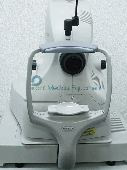
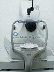
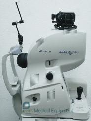
Topcon 3D OCT-2000 Optical coherence tomography sale Includes:
- OCT-2000 Unit
- Power Table
- Computer
- Fixation Light
- User's manual
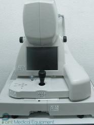
The Topcon 3D OCT-2000 System is the first Spectral Domain OCT system to incorporate a high resolution fundus camera and a user friendly color touch screen display in a compact, space saving design. The easy-to-use, intuitive FastMap™ software enables dynamic viewing of the OCT data, providing 3D, 2D and fundus images simultaneously. Pin-Point™ Registration properly indicates the location of the OCT image within the fundus image. In addition, the compare function allows users to view serial exams in a comparison view and apply different analytical tools. The seamless integration of the 3D OCT-2000 with Topcon's EyeRoute® Image Management System provides true connectivity and access to images anywhere, anytime.
What makes the Topcon 3D OCT-2000 unique:
- Integrated, high resolution (12.3MP) fundus camera
- FastMap™ software enables dynamic viewing of 2D, 3D and fundus images simultaneously
- Embedded touch-screen for quick and easy navigation
- Historic patient data from Stratus® OCT can be easily imported, analyzed and viewed
- Seamless integration with EyeRoute® Image Management System
High Quality Imaging
- 3D OCT Image: Topcon’s proprietary FastMap software pioneered the 3D visualization of OCT data, providing another dimension of clinical information, thereby enhancingthe understanding and illustration of complex pathologies such as vitreous traction, macularedema and retina schesis.
- 3D visualization of OCT data
- Illustrates complex pathologies
- Quickly export images and 3D movies for presentations
- Fundus Image: High resolution, non-mydriatic, color fundus images allow for visualization of conditions which otherwise would go undetected with OCT technology, such as disc hemorrhages. The 45 degreefield of view and the availability of stereo photos provides the ultimate diagnostic insight.
- High resolution 12.3 MP color fundus camera
- Easily capture and view stereo photos
- PinPoint Registration™ of OCT data in the fundus image
- B-scan Image: FastMap software encompasses the latest in noise reduction algorithms and overlapping scanning technology to create exquisite B-Scan images, which are available almost instantaneously. Thisgreatly reduces chair time for the patient and enhances office workflow.
- Uses latest in noise reduction algorithms
- Enhances office workflow
- Customize capture protocols
Deeper Diagnostic Insights
- Comprehensive Capturing: Capture images of the fovea and optic nerve head in one singlescan and high resolution images of the choroid with automatic choroid reference mode.
- Video Functions: Use single touch control to review and playback images and create 2D and 3D videos.
- Extended Scanning Depth: Capture high-quality images of high-myopic and hyperopic patients with a diopter compensation lens and an extended scanning depth of 2.3 mm.
- Compare Function: Allows you to visualize serial exams or view both eyes side by side
- FastMap Software: Topcon’s proprietary FastMap software ensures consistent, high quality images. Using enhanced 3D registration technology, FastMap reduces artifacts which may be caused by eye movement. This in turn allows for speedy PinPoint Registration of the OCT data within the fundus image without sacrificing workflow and preserves image quality of the vitreo-retinal interface. In addition, FastMap provides dynamic, simultaneous viewing of the fundus image, and 2D and 3D OCT data, and automatically detects ILM, RFNL, IS/OS junction, RPE and Bruch’s membrane, which can also be modified.
- Thickness Measurement Functions: FastMap software incorporates the latest layer detection algorithms, allowing you to automatically measure total retinal thickness, RNFL or compare against your legacy Stratus measurements. Manual adjustment of all measurement grids combined with auto and manual registration of serial exams gives you the highest level of confidence in retinal and RFNL thickness measurements.
- Mosaic Function: Create panoramic views from the macula to the optic disc.
Resources: Topcon_OCT_2000_Brochure.pdf