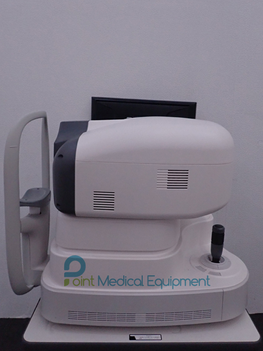

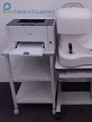
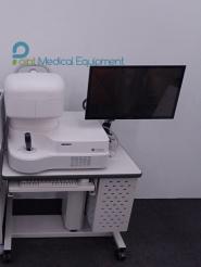

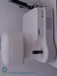
Tomey CASIA 2 Anterior ocular segment 3D OCT Package sale includes:
The Tomey CASIA2 provides an impressive user experience with intuitive operation and automation, supported by an unbelievable measurement speed. Our software guides you through measurement, analysis and the final report. Get inspired now and see the eye from a different perspective
Excellent features
OCT (anterior ocular segment three dimensional optical coherence tomography) anterior ocular segment. This OCT anterior ocular segment which enables non-contact/ non-invasive 3D anterior segment imaging. State-of-the-art inspection system that measures the cross-sectional image up to the lens and the shape of the cornea with an inspection method with little risk of infection.
Cornea Application
A topographic image is taken only in 0.3 seconds, providing high-quality images with no artifacts. The cornea can be displayed in different map types in a wide range of individual settings, either in 10 mm or 16 mm diameter. Scleral lens fitting is much easier than before.
Epithelium Map Application
With the new detailed epithelium map,* also available in sectors, the topography function is now more enhanced and provides advanced support for your treatment
STAR 360° & Glaucoma Application
With our STAR 360° application, the CASIA2 automatically measures the anterior chamber angles all around the anterior segment, thanks to its automatic scleral spur detection, and it provides you with specific paramaters needed to detect and treat glaucoma. With the new* added function ”Narrow Angle Index” you immediately receive data about a possible narrow angle plus a referring index based on normative data. *available from version 60.
Cataract Application
The special Pre-OP Cataract application guides you towards the ideal preparation for your cataract surgery. It displays essential and necessary screening parameters for your preferred IOL choice. The Post-OP application clearly visualises and documents the quality of your cataract surgery and provides you more information evaluating the outcome of your cataract surgery. The new colour camera enhances the check for toric IOL’s by seeing the toric markers in different options for correct positioning of your toric lens. Furthermore, a new scan type is added for the Post-OP Cataract application (IOLSCAN), which enhances the view on the IOL tremendously.
Total Analysis
Anterior segment images and parameters of both eyes can be verified at a glance. In addition to the conventional pre- and post-operative exam inations, CASIA2 can be used for daily clinical operation such as initial examination, follow-up treatment and obtaining informed consent. Each parameter is colour-coded to call attention if there is a difference between the left and right eye, or if it is within a specified range.
Pre-OP ICL
With two different integrated size recommendation formulas, this application enables the doctor to confirm the correct sizing for ICL surgery.
Post-OP ICL
The new Post-OP ICL movie function* controls the dynamic vault and shows the vault under mydriasis and miosis conditions as well as the relevant vault range difference. A fantastic tool for a proper quality check.
*available from version 60
Reference study: ”Dynamic Assessment of Light Induced Vaulting Changes of Implantable Collamer Lens with Central Port by Swept-Source OCT: Pilot Study” Dr. Felix Gonzalez-Lopez; Madrid, Spain.
| Measuring Mode 3D Mode | ||
| Anterior segment (Standard) | Scan direction | Radial Scan – 128 images |
| Scan resolution | 512 lines A scan per image sampling | |
| Scan speed | 2.4 sec | |
| Scan range (cross-section) | 16mm | |
| Angle analysis | Scan direction | Radial scan – 128 images |
| Scan resolution | 512 lines A scan per image sampling | |
| Scan speed | 2.4 seg | |
| Scan range (Cross-section) | 16mm | |
| Angle HD | Scan direction | Raster scan Horiz/Vert – 64 images |
| Scan resolution | 512 lines A scan per image sampling | |
| Scan speed | 1.2 seg | |
| Scan range (Cross-section) | 8mm X 4mm | |
| Bleb | Scan direction | Raster Scan Horiz/Vert – 256 images |
| Scan resolution | 256 lines A scan per image sampling | |
| Scan speed | 2.4 seg | |
| Scan range (Cross-section) | 8mm X 8mm / 12mm X 12mm | |
| Corneal Map | Scan direction | Radial scan – 16 images |
| Scan resolution | 512 lines A scan per image sampling | |
| Scan speed | 0.3 sec | |
| Scan range(Cross-section) | 10mm | |
| 2D Mode | ||
| Anterior Segment (Standard) | Scan resolution | 2048 lines A scan per image sampling |
| Scan range (Cross-section) | 16mm X 16mm | |
| Angle analysis | Scan resolution | 2048 lines A scan per image sampling |
| Scan range (Cross-section) | 16mm X 16mm | |
| Angle HD | Scan resolution | 2048 lines A scan per image sampling |
| Scan range (Cross-section) | 8mm X 8mm | |
| Movie mode | ||
| Anterior segment (Standard) | Scan resolution | 512 lines A scan per image sampling |
| Scan range (Cross-section) | 16mm X 16mm | |
| Angle analysis | Scan resolution | 512 lines A scan per image sampling |
| Scan range (Cross-section) | 16mm X 16mm | |
| Angle HD | Scan resolution | 512 lines A scan per image sampling |
| Scan range (Cross-section) | 8mm X 8mm |
Download