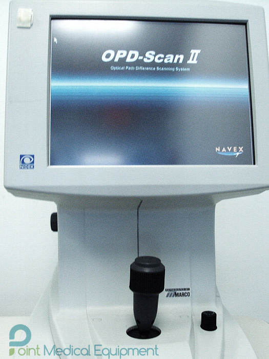

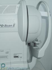
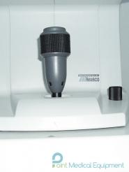
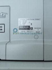
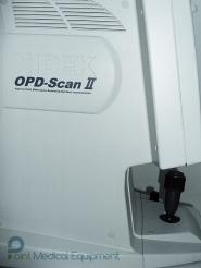
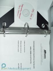
Item only includes Nidek OPD-Scan II Marco ARK 10000 Autorefractor Topographer
* Excellent Condition
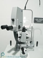
Nidek OPD-Scan II Marco ARK 10000 Autorefractor Topographer provides information on corneal topography, wavefront, autorefraction, keratometry and pupillometry in one unit. Ultilizing state-of-the-art imaging and analysis technology developed specifically to measure normal to high aberrated eyes.
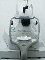
Nidek OPD-Scan II Marco ARK 10000 Autorefractor Topographer measures the refractive condition and analyzes corneal shape of the patient’s eye.
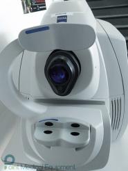
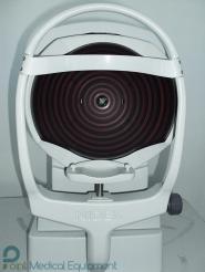
The refractive condition is measured by weak infrared rays, and corneal shape is analyzedbased on projected placido rings onto the cornea.
Accurate and Reliable Data for Optic Diagnostics
The NIDEK OPD-Scan II provides information on corneal topography, wavefront, autorefraction, keratometry and pupillometry in one unit, utilizing state-of-the-art imaging and analysis technology developed specifically to measure normal to highly aberrated eyes. The system offers a variety of data maps to provide information on the total refractive error, wavefront, corneal shape, internal aberrations and visual quality of the eye, allowing highly accurate and reliable information for optic diagnostics
The OPD-Scan II utilizes the principle of skiascopic phase difference for refractive error map measurement. The retina is scanned with an infrared light slit beam, and the reflected light is captured by an array of rotating photo detectors over a 360º area
The corneal topography function utilizes Placido disc technology. The system captures the image of reflected rings of light from the cornea and analyzes thousands of data points to plot the corneal contour, shape and refractive power
The OPD-Scan II measures corneal refractive power by corneal topography, and total refractive error as the OPD map. The Internal OPD Map plot is created by subtracting the corneal refractive power from the total OPD to display in diopters the distribution of refractive error contained in the internal eye
Resources: